
howitreat.in
A user-friendly, frequently updated reference guide that aligns with international guidelines and protocols.
Hematopoiesis
Definition:
- It is the term used to describe the formation and development of blood cells.
- Hemangioblasts give rise to both circulatory system and blood cells. They are derived from mesoderm.
Stages and sites:
- Primitive hematopoiesis
- Starts in aorto-gonado-mesonephros region of embryo at 19th day after fertilization.
- It is called “blood island”, which consists of hemocytoblasts and erythroblastoid cells surrounded by endothelial cells.
- This continues up to 7 weeks of gestation.
- Hemoglobins of embryonic variety- Gower 1, Gower 2 and Portland are produced.
- Hepatic erythropoiesis
- Starts in 3rd month and continues up to 6 months of gestation
- Between 7 and 15 weeks, 60% of liver cells are of hematopoietic origin.
- HbF is the type of hemoglobin produced during this phase.
- During this phase, to some extent, spleen, thymus and kidney and lymph nodes also show hematopoiesis.
- Medullary (Bone marrow) hematopoiesis
- Starts at 3rd trimester of gestation
- Produces HbA and HbA2
- At birth throughout the skeleton, there is hematopoiesis.
- In adults, hematopoiesis occurs, mostly in sternum, pelvis, skull, ends of long bones and vertebra.
Stem cells:
- They are undifferentiated cells, capable of dividing for indefinite periods, to self-renew and to generate functional progeny of highly specialized cells.
- Hierarchy of stem cells
- Totipotent (Present in zygote)
- Pluripotent (Present in embryo)
- Multipotent (Adult tissues such as bone marrow)
- Oligopotent
- Unipotent
- 1013 new cells are needed every day to maintain steady state of blood counts (i.e., 6 billion cells/kg/day).
- There is one stem cell per 20 million nucleated cells.
- Stem cell pool consists of 1-2x10⁶ cells
- When a stem cell divides, one cell remains in the stem cell pool, while the other cell goes into maturation pathway.
- Stem cell plasticity- It is the ability of the hematopoietic and stromal stem cells to produce cells associated with other tissues, such as liver, lung and muscle. This differentiation depends on the cytokines present in the micro-environment.
- Stem cell trafficking- It is the movement of stem cells from bone marrow into circulation and “homing in” of circulating stem cells into the bone marrow. Normally very few of stem cells are released into peripheral blood and most of them return back and home in.
- “Homing in” involves transendothelial migration from the blood stream into the marrow micro-environment. Chemokine- CXCL-12, the ligand for CXCR-4 expressed on stem cells, plays important role in homing in.
- Mobilization- It is detachment of stem cells from the micro-environment and transendothelial migration in the reverse direction. Matrix metalloproteases and reactive oxygen species, which are produced during G-CSF administration, degrade the components of endothelial tight junctions and enhance stem cell egress.
- Immunophenotyping of stem cells
- Positive- CD34, CD90, CD133, CD117, CXCR4, Scal-1, CD164, CD150, CD110
- Negative- CD33, CD38, HLA-DR, Lineage specific markers
- Anti-CD 34 antibodies, tagged with magnetic beads can be used for isolation of stem cells. This process is called as cell sorting. 1% of human bone marrow cells express CD34.
- Stem cells are resistant to chemotherapy induced cytotoxicity due to high level of expression of drug efflux pumps (verapimil sensitive)
Erythropoiesis:
| Stage | Size (microns) | Nucleus | Cytoplasm |
| Pronormoblast | 12-20 | Large, round, with several nucleoli | Deep and basophilic |
| Early normoblast | 10-16 | Large coarse chromatin, no nucleoli | Basophilic |
| Intermediate normoblast | 8-14 | Smaller nucleus, coarse chromatin which is deeply stained | Hemoglobin starts appearing, hence acidophilic tint. |
| Late normoblast | 8-10 | Small and pyknotic | Acidophilic |
| Reticulocyte | 8 | No nucleus | Diffuse pale basophilia |
| Red blood cell | 7-7.5 | No nucleus | Completely hemoglonized |

Figure 1.1.1. - Pronormoblast (Single arrow) and early normoblast (Double arrow)
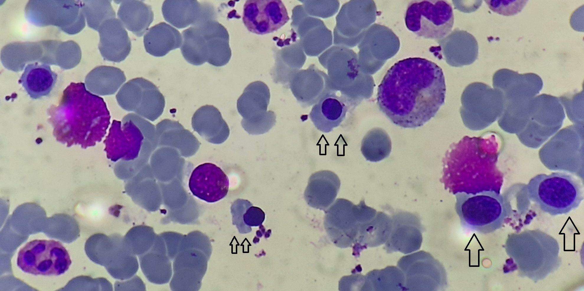
Figure 1.1.2. - Intermediate normoblast (Single arrow) and late normoblast (Double arrow)
- Within bone marrow, erythroblasts are organized into erythroblastic islands which contain central 1-2 macrophages, surrounded by layer of erythroblasts in different stages of development.
- Hemoglobin content has a negative feedback on cell proliferation and differentiation
- If hemoglobin content is less (as in iron deficiency), cells undergo extra division, yielding smaller hypochromatic cells.
- If hemoglobin synthesis exceeds DNA synthesis (as in case of megaloblastic anemia), cells skip a division and nuclear extrusion occurs early, leading to macrocytosis.
- Rate of erythropoiesis can increase to 5-10 folds in response to appropriate erythropoietin stimulation, if sufficient iron store is present.
Regulation of erythropoiesis
Hypoxia
↓
Lack of oxygen for ferrous iron propylhydroxylase enzyme
↓
No hydroxylation of proline residues of HIF-1-alpha, hence no degradation of HIF-1-alpha (Normally this happens through ubiquitation by von Hippel Lindau protein)
↓
HIF-1-alpha heterodimerizes with HIF-1-beta which is constitutively present
↓
Activation of following hypoxia responsive genes, resulting in increased production of various factors such as
Erythropoietin- Binding of EPO to EPO receptor induces conformational change in EPO receptor, which activates JAK-2 pathway, which stimulates proliferation of erythroid cells.
Vascular endothelial growth factor
Transferrin receptors
Granulopoiesis:
| Stage | Size (microns) | Nucleus | Cytoplasm |
| Myeloblast | 10-18 | Round to oval, eccentric/central, evenly stained chromatin with reticular (fine meshwork) appearance, 1-6 nucleoli | Scanty, bluish, Few or no granules |
| Promyelocyte | 12-20 | Round to oval, eccentric/central, Nuclear chromatin is condensed and nucleoli are less well defined | Moderate amount, deep blue, some azurophilic granules are present |
| Myelocyte | 12-18 | Oval/slightly indented, Eccentric, No nucleoli, thick, deeply stained coarse chromatin, rarely nucleoli seen | Moderate amount, Bluish pink, Specific granules are present, pale pink cytoplasm |
| Metamyelocyte | 10-18 | Thick horse shoe/ indented, Central/eccentric, Smaller and indented, No nucleoli | Plentiful amount, Pink cytoplasm with granules. |
| Band forms | 10-16 | Band shape of uniform thickness, Central/eccentric, Has deep indentation and coarsely clumped chromatin, No nucleoli | Plentiful amount, Pink, Plenty of granules. |
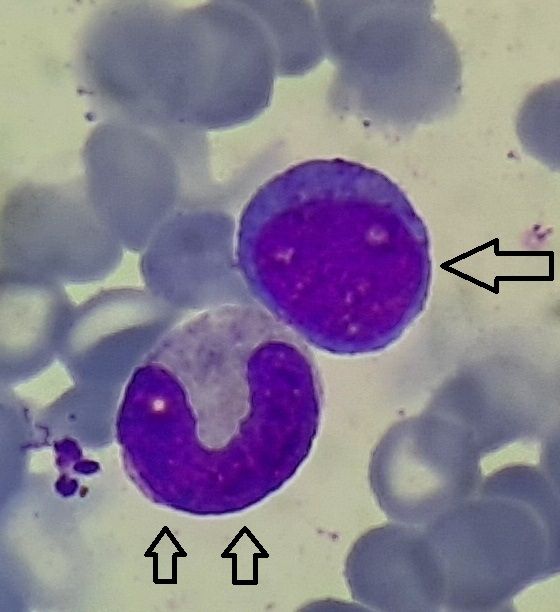
Figure1.1.3 - Myeloblast (Single arrow), Band form (Double arrow)
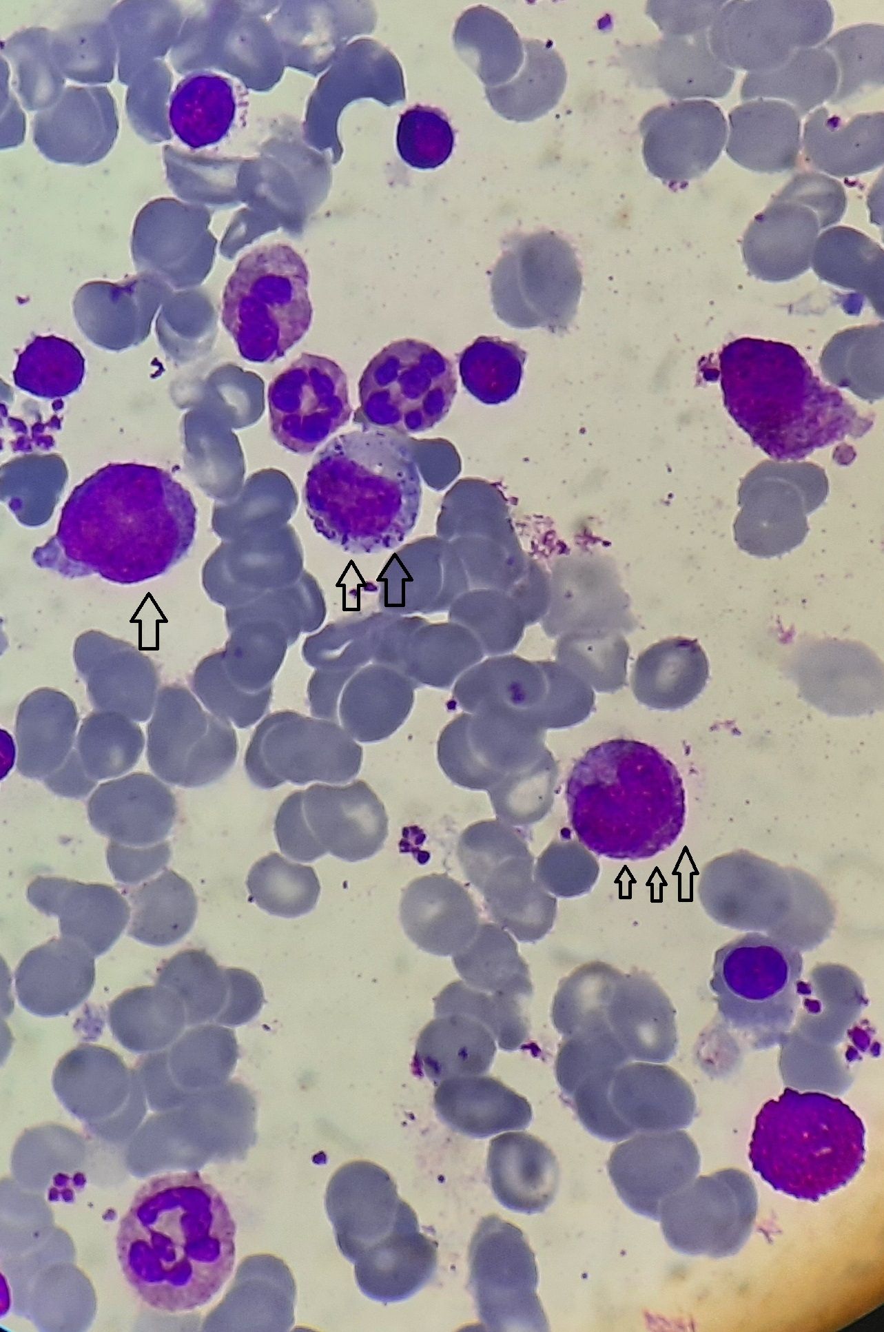
Figure 1.1.4 - Promyelocyte (Single arrow), Myelocyte (double arrow), Metamyelocyte (Triple arrow)
Morphology of each type of mature granulocyte is explained in WBC section.
Monocytic series
| Stage | Size (microns) | Nucleus | Cytoplasm |
| Monoblast | Similar to myeloblast | Similar to myeloblast | Similar to myeloblast |
| Promonocyte | 12-20 | Eccentric/ central, Large convoluted, chromatin in skein like strands, 1-2 nucleoli | Moderate amount, Dull gray blue with few azurophilic granules |
| Monocyte | 12-20 | Eccentric/ central, round/oval/horse shoe shaped, chromatin in skein like strands, no nucleoli | Abundant, grayish, with numerous, fine lilac granules |
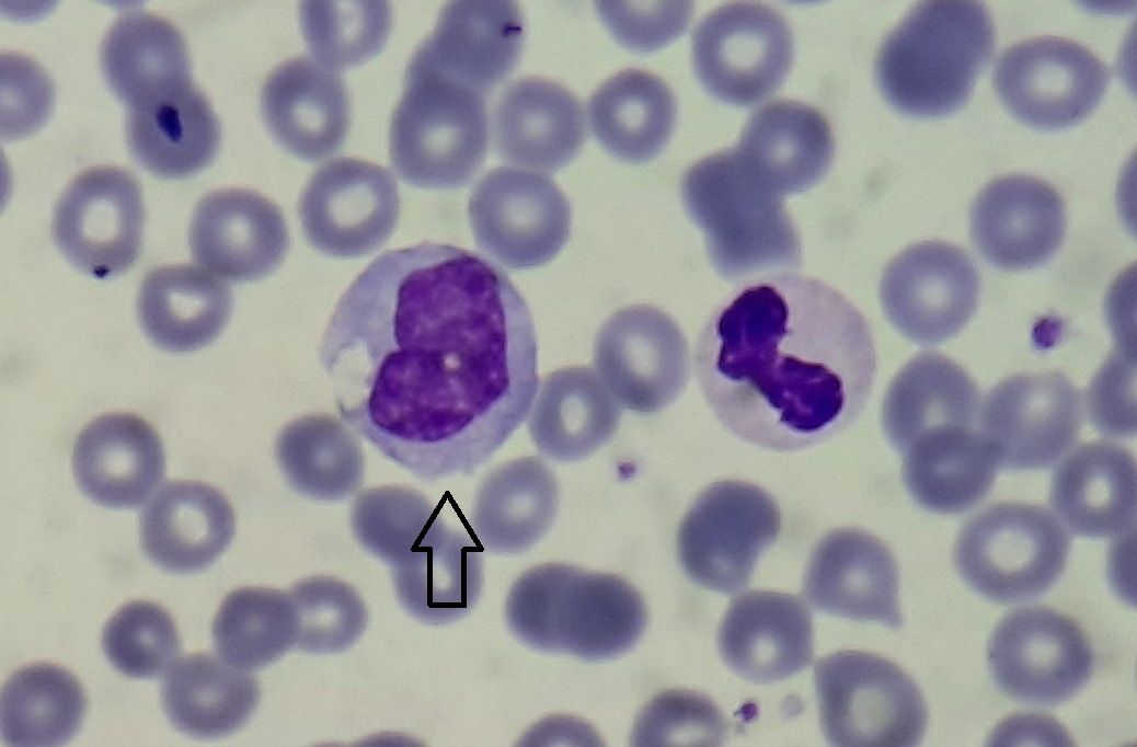
Figure1.1.5 - Monocyte (Arrow)
Lymphopoiesis:
| Stage | Size (microns) | Nucleus | Cytoplasm |
| Lymphoblast | 15-20 | Eccentric/central, round/oval, Chromatin has fine reticular appearance, has up to 2 nucleoli | Agranular, basophilic |
| Large lymphocyte | 12-15 | Round with clumped chromatin | Pale sky blue |
| Small lymphocyte | 8-10 | Round nucleus, heavily clumped chromatin. | Grayish, clear |
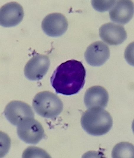
Figure 1.1.6 - Lymphocyte
Megakaryopoiesis:
| Stage | Size (microns) | Nucleus | Cytoplasm |
| Megakaryoblast | 20-30 | Large, oval/ kidney shaped, several nucleoli | Scanty, agranular |
| Promegakaryocyte | 14-30 | Mild lobulations, denser chromatin | Intensely basophilic with fine azurophilic granules |
| Megakaryocyte | 30-90 | Single, multilobulated nucleus (4-16 lobes) | Bulky, light blue with fine azurophilic granulations. Margin is irregular with budding. |
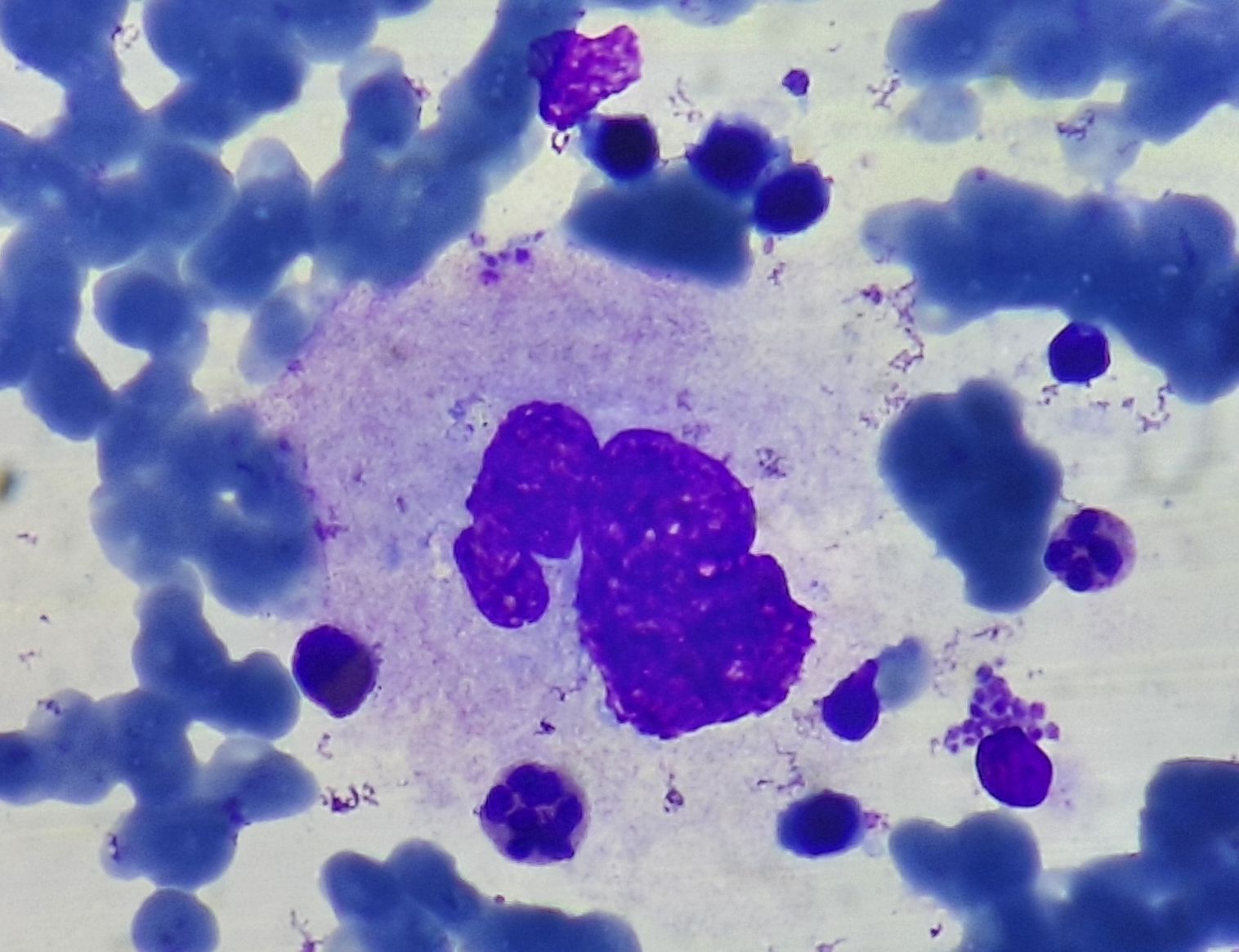
Figure 1.1.7- Promegakryocyte
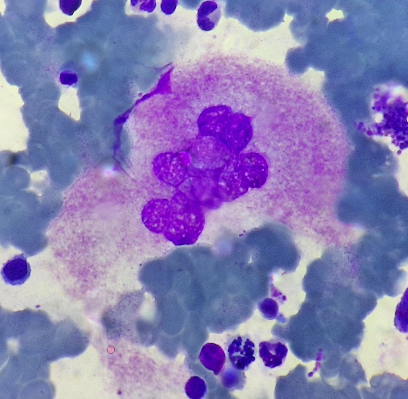
Figure 1.1.8- Megakryocyte
- Megakaryocytes are 1micrometer away from BM sinus and shed platelets directly into the sinus.
- Platelets are released in groups called proplatelets, which later on break to form platelets. This breaking down principally occurs in the lung.
- 5 days are needed for formation of platelets from megakaryocytes.
- Each day 1011 platelets are produced.
- Life span of platelet is 7-10 days
- Each megakaryocyte gives rise to 1000 to 3000 platelets before the residual nuclear material is engulfed and eliminated by marrow macrophage.
- Nuclear maturation takes place first and largely complete before cytoplasmic maturation starts.
- Nuclear maturation consists of series of endomitosis, in which the DNA content of the cell is doubled but cell division does not take place. So, ploidy becomes 4N to 128N.
- IL6, IL11 and thrombopoietin stimulate megakaryocyte proliferation and differentiation.
- Thrombopoietin is produced in liver and it acts through c-Mpl receptor on megakaryocytes.
- Platelets also have c-Mpl receptors, which bind and clear TPO from circulation.
- Emperipolesis- In marrow sections, neutrophils or other marrow cells are seen transiting through the cytoplasm of mature megakaryocyte. This is called emperipolesis. There is no pathological significance for this.
Cytokines and genes involved in hematopoiesis
(Upregulated genes are written in bracket)
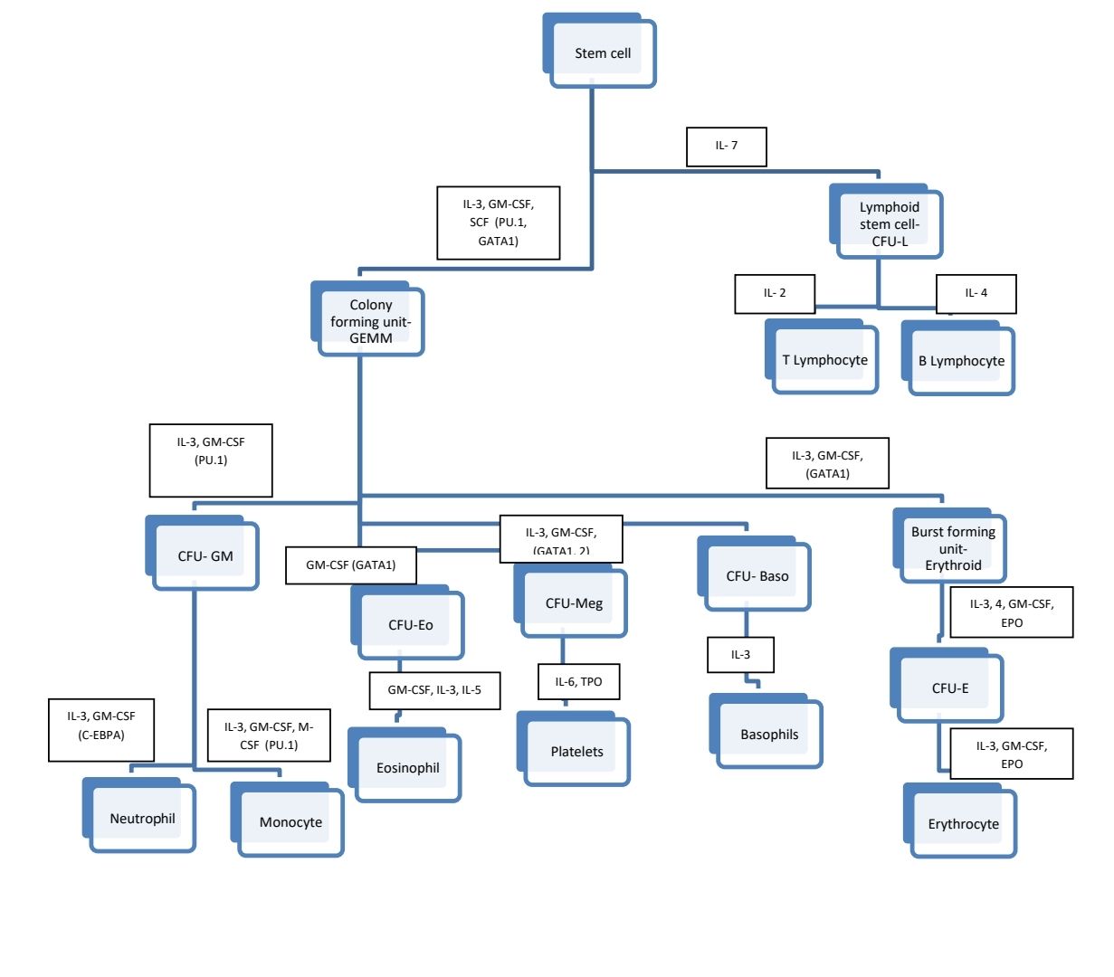
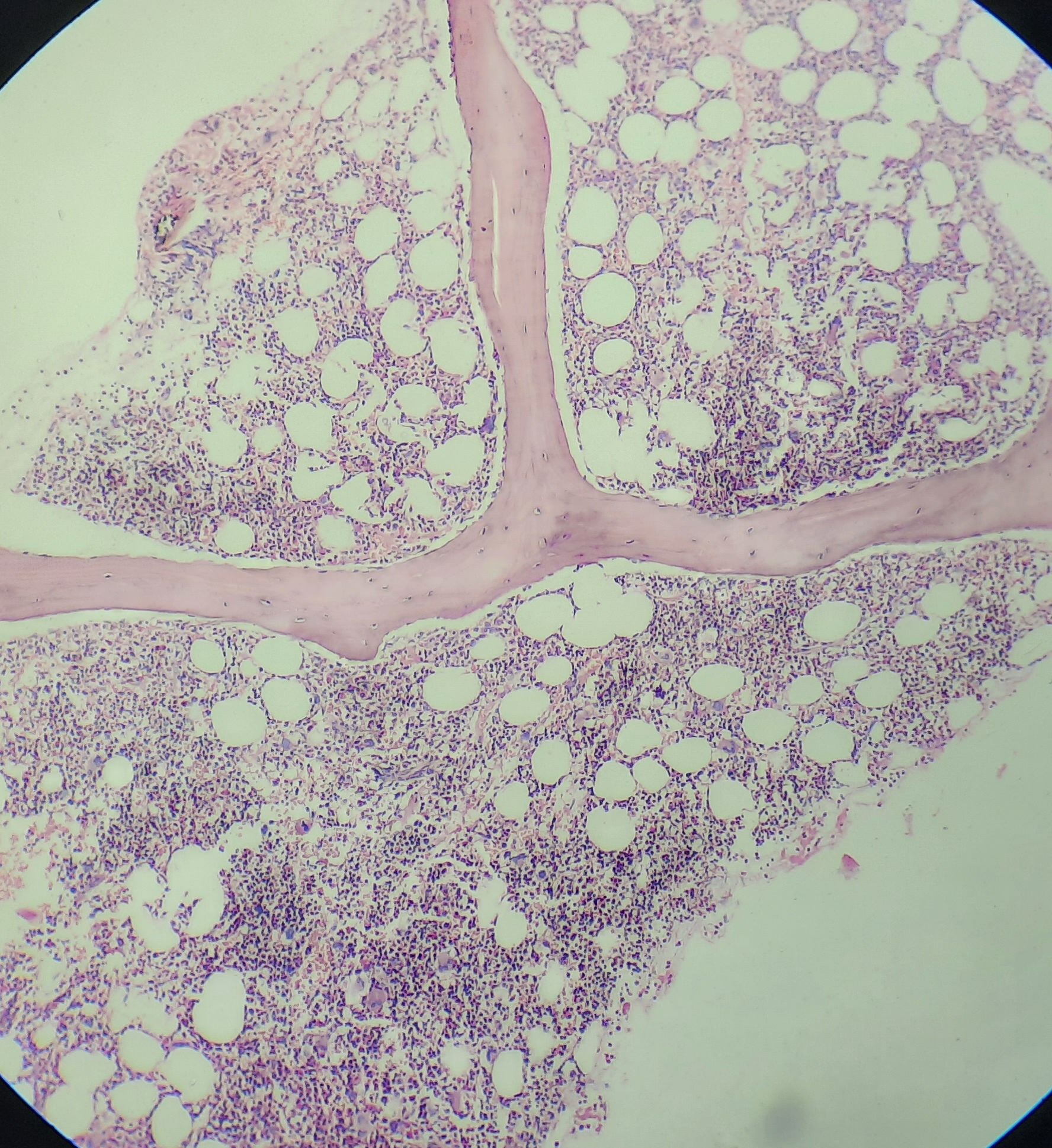
Figure 1.1.9 - Normal trephine biopsy of a young person
Bone marrow microenvironment: It consists of following elements
- Marrow stroma
- Sinusoids lined by endothelial cells and surrounded by adventitial reticular cells. Latter cells are fibroblasts which are capable of transforming in to adipocytes.
- Innervation- Both myelinated and non-myelinated nerve fibers are present in the periarterial sheath of marrow. Non-myelinated nerve endings secrete neurohumors that affect hematopoiesis.
- Osteoblasts and osteocytes
- Osteoclasts
- Mesenchymal stem cells- Located in the perisinusoidal space.
- Macrophages
- Lymphocytes
- Plasma cells
- Extracellular matrix- Composed of proteoglycans (Heparin sulphate, chondroitin sulphate), fibronectin, tenacin, collagen, laminin, hemonectin, thrombospondin, vitronectin
- Cell adhesion molecules
- Integrins
- Immunoglobulins
- Selectins
- Sialomucins
- Hyaluridin
- Various cytokines such as G-CSF, GM-CSF, glucocorticoids etc.
An Initiative of
Veenadhare Edutech Private Limited
1299, 2nd Floor, Shanta Nivas,
Beside Hotel Swan Inn, Off J.M.Road, Shivajinagar
Pune - 411005
Maharashtra – India

howitreat.in
CIN: U85190PN2022PTC210569
Email: admin@howitreat.in
Disclaimer: Information provided on this website is only for medical education purposes and not intended as medical advice. Although authors have made every effort to provide up-to-date information, the recommendations should not be considered standard of care. Responsibility for patient care resides with the doctors on the basis of their professional license, experience, and knowledge of the individual patient. For full prescribing information, including indications, contraindications, warnings, precautions, and adverse effects, please refer to the approved product label. Neither the authors nor publisher shall be liable or responsible for any loss or adverse effects allegedly arising from any information or suggestion on this website. This website is written for use of healthcare professionals only; hence person other than healthcare workers is advised to refrain from reading the content of this website.