
howitreat.in
A user-friendly, frequently updated reference guide that aligns with international guidelines and protocols.
Inherited Bone Marrow Failure Syndromes
Introduction:
- These are the conditions where in there is decreased production of one or more of the major hematopoietic lineages due to mutations that were derived from parents or occurred de-novo.
- These mutations directly affect physiological cell survival and function pathways that are essential for normal hematopoiesis, such as
- DNA repair
- Telomere maintenance
- Ribosome biosynthesis
- Microtubule stabilization
- Signaling from hematopoietic growth factors
- Signal transduction related to hematopoietic cell differentiation
Classification:
- IBMFSs with pancytopenia
- Fanconi anemia
- Schwachman Diamond syndrome
- Dyskeratosiscongenita
- Pearson syndrome
- Reticular dysgenesis
- Lig4-associated aplastic anemia
- IBMFSs with predominantly anemia
- Diamond Blackfan anemia (Refer: Pure red cell aplasia)
- Inherited sideroblastic anemia (Refer: Sideroblastic anemia)
- Congenital dyserythropoietic anemia (Discussed in separate chapter)
- IBMFSs with predominantly neutropenia (Refer: Miscellaneous diseases section)
- Kostman syndrome
- Dursun syndrome
- Cyclic neutropenia
- WHIM syndrome
- Glycogen storage disease Ib
- Barth syndrome
- Poikiloderma with neutropenia
- IBMFSs with predominantly thrombocytopenia (Refer: Platelet disorders section)
- Congenital amegakaryocytic thrombocytopenia
- Thrombocytopenia absent radii
- Epstein, Fechtner, Sebastian, May Hegglin, Alport syndrome
- Mediterranean platelet disorder
- Familial autosomal dominant nonsyndromic thrombocytopenia
- Thrombocytopenia with dyserythropoiesis
- Thrombocytopenia with associated myeloid malignancies
- X linked thrombocytopenia
- Thrombocytopenia with radio-ulnarsynostosis
Fanconi Anemia
- It is inherited in autosomal recessive pattern
- Incidence: 1-5cases / million
- Genes affected include.
Complementation Group/ | Approximate percentage of FA patients | Chromosome location | Protein (amino acids) | Mutations identified |
A (FANCA) | 65-70 | 16q24.3 | 1455 | >120 |
B (FANCB) | <2 | ? | ? | ? |
C (FANCC) | 5-10 | 9q22.3 | 558 | 10 |
D1(FANCD1) | < 2 | 13q12-13 | 3417 | 9 |
D2 (FANCD 2) | < 2 | 3p25.3 | 1451 | 5 |
E (FANCE) | 2-5 | 6p21.3 | 536 | 3 |
F (FANCF) | < 2 | 11p15 | 374 | 6 |
G (FANCG) | 10-15 | 9p13 | 622 | 21 |
Many others have been identified.
Pathogenesis:
- Nuclear complex containing FA proteins (A/C/E/F/G) is required for activation of FANCD2 protein to monoubiquitinatedisoform
- In normal cells FANCD2 is monoubiquitinated at lysine 561 in response to DNA damage (e.g.-MMC) & is targeted to discrete nuclear foci
- FA genes act along with BRCA1 & BRCA 2 genes to bring about DNA repair (Called as FA/BRCA pathway)
In Fanconi anemia hematopoietic progenitors are hypersensitive to TNF-α & INF-γ
↓
DNA damage and defective repair
↓
Increased Fas induced apoptosis
- 90% of affected people develop aplastic anemia.
- Pancytopenia appears at 5-10 years of age.
- There is increased incidence of development of malignancies such as AML (especially M4 & M5), hepatic tumors, brain tumor, Wilm tumor and squamous cell carcinoma
Physical Findings Associated with Fanconi Anemia
- Skeletal
- Short stature
- Radial anomalies (e.g. thumbs, hands, and forearms)- Hypoplastic/ supernumerary/bifid/absent thumbs
- Microcephaly
- Hip and spine anomalies
- Toe anomalies: syndactyly, short toes, supernumerary toes, club foot and flat foot
- Skin
- Hyperpigmentation (e.g., café au lait spots)
- Hypopigmentation (e.g., vitiligo)
- Genitourinary
- Renal anomalies – Hypoplasia of kidney, ectopic/ horseshoe kidney
- Hypogonadism- Underdeveloped penis, undescended/atrophic/absent testis, Hypospedias, phimosis, abnormal urethra, malformations of vagina, uterus and ovaries
- Craniofacial
- Ophthalmic anomalies (e.g. microphthalmia and epicanthal folds)
- Otic anomalies (e.g. external and internal ear anomalies and deafness)
- Gastrointestinal malformations – Anorectal and duodenal atresia
- Cardiac malformations
Investigations:
- Peripheral smear- Macrocytic anemia, later pancytopenia once marrow aplasia develops
- Stress karyotyping on peripheral blood lymphocyte cultures/ cultured skin fibroblasts
- Addition of diepoxybutane and mitomycin C to metaphase preparatio
- Karyotype shows increased chromosomal breaks, gaps, rearrangements, exchanges and reduplications
- Triradiate chromosomes and chromosomal breaks are seen even in patients who have not yet developed aplasia
- Do not use bone marrow samples because of false negative results
- S. Alpha feto protein- Increased
- HbF- Increased
- Ubiquitation of FANCD2
- Sequencing and identification of mutation
- USG and Echo- for internal organ anomalies
- BM Aspiration-
- Early stage- shows erythroid hyperplasia, sometimes with dyserythropoiesis, myelodysplastic changes or megaloblastic changes
- Later stages- Hypocellular bone marrow with relative increase in lymphocytes, plasma cells and mast cells
Prognosis
- Median survival – 24 years
Treatment
- For patients who do not have transfusion requirement, period of observation is indicated. Monitor CBC once in 3months and BM aspiration annually in these patients. Also do surveillance for solid tumors every year.
- Surgery for correction of hand deformity (Index finger is placed at the position of thumb). This should be done before 2 years of age. Otherwise brain cannot recognize index finger as thumb.
- Improve hematopoietic function by
- Androgens
- PO. Oxymetholone- 1-5mg/kg- OD or IM. Nandrolonedecanote- 1-2mg/kg/week or Danazol
- Overall response rate- 50%
- Overall response time- 1-2 months
- Once a maximal response is achieved slowly taper the androgens
- Indications- Hemoglobin <8gm/dL, Platelet count- <30,000/cmm, ANC <500/cmm
- Eventually patients become refractory and bone marrow failure progresses
- Corticosteroids – Prednisolone
- G-CSF- Useful in case of neutropenia
- Androgens
- Hematopoietic stem cell transplantation (Treatment of choice)
- Indications:
- Severe underproductive cytopenia and transfusion dependency
- High risk MDS with chromosomal clonal abnormalities like monosomy 7, partial trisomies and tetrasomies
- Overt AML
- Caveats
- More than 20 exposures to blood products is a risk factor that adversely affects engraftment and survival post-transplant
- Use of directed donations from family members can cause allo-immunization
- It should be done before the onset of MDS/AML and before multiple blood transfusions are given
- Sibling donor should be tested with thorough history, examination, blood counts, HbF and chromosomal breakage studies to exclude Fanconi anemia in them.
- As Fanconi anemia cells are hypersensitive to radiation and chemotherapy, pre-stem cell transplantation conditioning is modified by reducing the dose. Ex: Low dose cyclophosphamide – 20mg/kg with 4.5 – 6 Gy of thoraco abdominal irradiation
- To decrease risk of irradiation, new protocols use Fludarabine/ ATG.
- 2 year survival after Stem cell transplantation
- HLA matched – 70%
- Unrelated – 20-40%
- Better survival rates with
- Younger patient age
- Fewer than 20 exposures to blood products
- Higher pretransplant platelet counts
- Absence of previous treatment with androgens
- Normal pretransplant liver function tests
- Use of fludarabine in cytoreductive regimens
- Limited malformations
- Recipient sero negativity for CMV
- Children cured by HSCT are at increased risk of solid tumors particularly squamous cell carcinoma of tongue.
- Indications:
- Retroviral mediated somatic gene therapy: Normal wild FANC gene is introduced into one stem cell which repopulates the bone marrow. This mosaic pattern induces hematological improvement.
Prevention:
- Pre-implantation genetic diagnosis
Dyskeratosis Congenita
It is a characterized by
- Bone marrow failure
- Abnormal skin pigmentation- Face, neck chest and arms
- Nail dystrophy- Longitudinal ridging, splitting, pterygium formation, complete nail loss
- Mucosal leukoplakia- Especially of tongue, conjunctival, anal, urethral, genital mucosa can be involved.
- Epiphora (excessive tears due to nasolacrymal duct obstruction), conjunctivitis, blepharitis, loss of eye lashes, strabismus, cataracts, optic atrophy
- Cognitive/developmental delay
- Pulmonary disease- idiopathic pulmonary fibrosis
- Short stature
- Dental caries/tooth loss
- Esophageal stricture
- Hair loss/gray hair/sparse eyelashes
- Hyperhidrosis
- Malignancy
- Intrauterine growth retardation
- Gastrointestinal disorders- Esophageal strictures, hepatomegaly, cirrhosis
- Ataxia
- Hypogonadism/undescended testes/ hypospadias
- Microcephaly
- Urethral stricture/phimosis
- Osteoporosis/aseptic necrosis/scoliosis
- Mandibular hypoplasia
- Deafness
Subtypes:
DC subtype | Approximate percentage of DC patients | Chromosome location | Gene product | Mutations identified |
X-linked recessive | 40 | Xq28 (DKC-1) | Dyskerin | 30 |
Autosomal dominant | 5 | 3q21-3q28 (TERC, TERT) | hTR | 6 |
Autosomal recessive | 50 | NOP10/NOLA3, NHP2/NOLA2 | ? | ? |
Etiopathogenesis
- Like Fanconi anemia, cells of Dyskeratosis congenita also display hypersensitivity to clastogenic agents like MMC, but rather than gaps/ breaks as in Fanconi anemia , there is chromosomal rearrangement.
- Skin fibroblasts in Dyskeratosiscongenita are abnormal both in morphology and in growth rate. They show unbalanced chromosomal rearrangements (dicentrics, Tricentrics, translocations) in the absence of any clastogenic agents (Peripheral blood & BM Metaphases also show similar changes)
- Hoyeraal – Hreidarsson syndrome is another syndrome due to mutation of same DKC 1 gene which is associated with
- Severe growth failure
- Abnormalities of brain development- cerebellar hypoplasia
- Aplastic anemia
- T+ B- NK- Severe combined immunodeficiency
- DKC 1 gene encodes protein Dyskerin, which is a nucleolar protein. It is involved in pseudouridylation of specific residues of r RNA
- Dyskerin also associates with RNA component of telomerase (hTR). Hence there is abnormal telomerase activity which leads to abnormally short telomeres for age.
Investigations:
- Telomere length: Shortened
- Hemogram: Macrocytic anemia, pancytopenia
- Bone marrow- Hypoplastic. (50% have severe aplastic anemia)
- Immunological abnormalities
- Decreased immunoglobulin levels
- Decreased T and B lymphocyte numbers
- Decreased or absent proliferative response to PHA
- MRI- Small sized cerebellum
Associated malignancies:
- Leukemia/ MDS
- Lymphoma
- Squamous cell carcinoma
- GI adenocarcinoma
- Lung/ Liver/ Skin carcinoma
Treatment
- Oxymetholone- 0.25-5mg/kg/day- Improves BM function in 50% cases. Danzol can be used as an alternative.
- G-CSF, GM-CSF, Erythropoietin
- Allogeneic stem cell transplantation: Increased sensitivity to transplant conditioning is observed which is related to telomere shortening. Hence low intensity, fludarabine based conditioning therapies are used
- Hematopoietic gene therapy
Revez syndrome:
- Dyskeratosis congenita with exudative retinopathy
- Autosomal dominant inheritance
- Due to mutation of TINF2 gene
Shwachman Diamond Syndrome
Etiology:
- Autosomal recessive condition
- Mutation of SBDS gene on chromosome 7q- It has important role in RNA metabolism/ ribosome biosynthesis
- Mutation leads to short telomeres and increased rate of apoptosis
Epidemiology:
- Incidence- 8.5cases/million live births
Clinical features:
- Exocrine pancreas insufficiency (100%)
- Bone marrow dysfunction (100%)
- Short stature (70%)
- Protuberant abdomen (60%)
- Icthyotic skin rash (60%)
- Metaphyseal dysostosis on X-ray (75%)
- Hepatomegaly
- Rib/thoracic cage abnormality
- Hypertelorism
- Synductyly
- Cleft palate
- Dental dysplasia
- Ptosis
- Skin pigmentation
- Hematological abnormalities
- Neutropenia (60%)
- Pancytopenia (20%)
- MDS
- Leukemic transformation (25%) – Most common is AML (M6)
Investigations:
- Bone marrow aspiration- Varying cellularity. Usually hypoplasia.
- Bone marrow cytogenetics:
- i(7q)- Seen in 44% of patients with MDS
- Monosomy 7
- Deletions and translocations involving chromosome 7q
- Pancreatic imaging: Extensive fatty replacement of pancreatic acinar tissue
- Serum trypsinogen: Decreased
- Skeletal imaging: Osteopenia, metaphyseal dysplasia, narrow rib cage, short ribs, digital abnormalities
- Brain imaging: Decreased global brain volume- Both gray and white matter, smaller posterior fossa, cerebellar vermis, corpus callosum, brainstem
Differential diagnosis: Other conditions with exocrine pancreatic insufficiency & hematological abnormality
- Pearson syndrome: It is associated with
- Anemia more prominent than neutropenia
- BM-Sideroblasts along with vacuolation of myeloid & erythroid precursors
- Acidosis
- Abnormalities of liver functions.
- Mitochondrial DNA rearrangements
- Poor prognosis- Most of them die before 5 years
- Cartilage hair syndrome
- Cystic fibrosis
Treatment:
- Oral pancreatic enzymes
- G-CSF-for neutropenia- But it may foster clonal evolution
- Oxymetholone
- Supportive treatment with Red cell transfusion, Platelet transfusion, and Antibiotics
- Bone marrow transplantation if there is
- Bone marrow failure with severe or symptomatic cytopenia
- MDS with excess of blasts/ Leukemia
Reticular dysgenesis
- It is a type of severe combined immunodeficiency
- X linked recessive with mitochondrial AK2 gene mutations
- Associated with lymphopenia, anemia and neutropenia
- Lymph nodes, tonsils and thymus are absent
- Treatment- Hematopoietic stem cell transplantation
Figures:
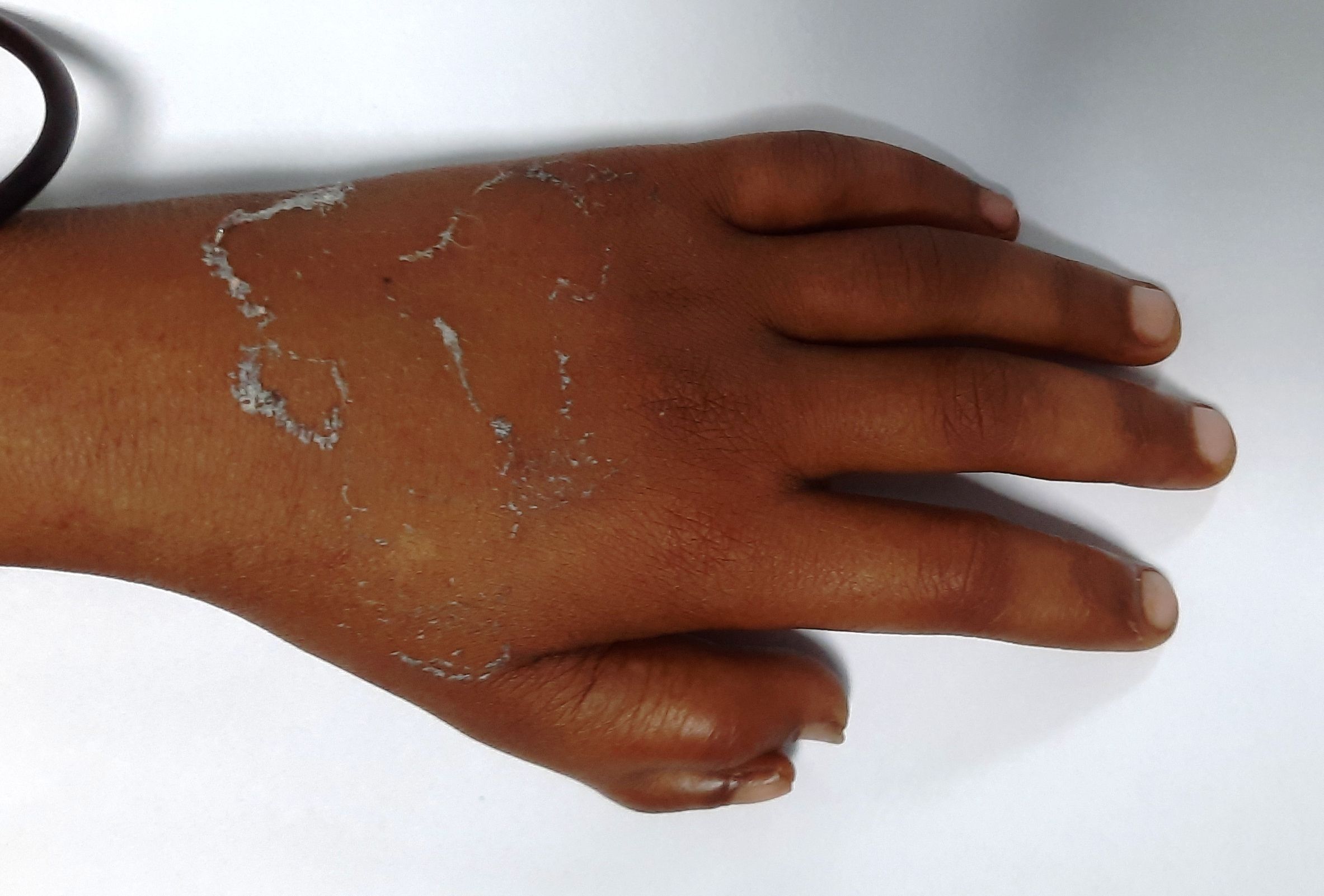
Figure 8.5.1- Thumb abnormality in Fanconi anemia
Recent advances:
Predictors of outcomes in hematopoietic cell transplantation for Fanconi anemia
The study retrospectively analyzed allogeneic hematopoietic cell transplantation (HCT) outcomes in Fanconi anemia (FA) patients (n=89) between 2007 and 2020. Overall survival (OS) at five years was 83.2%, and event-free survival (EFS) was 74%. Predictors for OS, EFS, and treatment-related mortality included age ≥19, HLA mismatch, and year of HCT. In the pediatric group, T-cell depletion (TCD) showed a borderline significance, with 5-year OS of 73.0% in TCD vs. 100% for T-replete HCT. The cumulative incidence of graft-versus-host disease and relapse was low, indicating excellent survival chances for FA patients undergoing HCT.
https://doi.org/10.1038/s41409-023-02121-1
HLA-haploidentical stem cell transplantation in children with inherited bone marrow failure syndromes
This retrospective study analyzed outcomes of haploidentical stem cell transplantation (haplo-SCT) in 162 children with inherited bone marrow failure syndromes (I-BMF), focusing on different T-cell depletion methods. Fanconi Anemia was the most common diagnosis (70.1%). Four T-cell depletion approaches were compared: TCRαβ+/CD19+ depletion, T-repleted with post-transplant Cyclophosphamide (PTCy), in-vivo T-depletion with ATG/alemtuzumab, and CD34+ positive selection. The study found that TCRαβ+/CD19+ depletion had lower incidences of acute and chronic GvHD and higher overall survival (79%) and GvHD/Rejection-free Survival (71%), highlighting its superiority for reducing severe GvHD and improving survival in I-BMF patients.
https://doi.org/10.1002/ajh.27293
Outcomes of hematopoietic stem cell transplantation in 813 pediatric patients with Fanconi anemia
A multicenter study of 813 pediatric patients with Fanconi anemia undergoing HSCT showed 5-year overall survival of 83%, with similar outcomes for matched family (88%) and unrelated donors (86%). Mismatched donors had lower survival rates (MMFD/MMUD: 72%; HID: 70%). Age ≥10 years and the presence of AML/MDS predicted worse outcomes. These findings suggest HSCT should be performed earlier in younger patients with well-matched donors.
https://doi.org/10.1182/blood.2023022751
An Initiative of
Veenadhare Edutech Private Limited
1299, 2nd Floor, Shanta Nivas,
Beside Hotel Swan Inn, Off J.M.Road, Shivajinagar
Pune - 411005
Maharashtra – India

howitreat.in
CIN: U85190PN2022PTC210569
Email: admin@howitreat.in
Disclaimer: Information provided on this website is only for medical education purposes and not intended as medical advice. Although authors have made every effort to provide up-to-date information, the recommendations should not be considered standard of care. Responsibility for patient care resides with the doctors on the basis of their professional license, experience, and knowledge of the individual patient. For full prescribing information, including indications, contraindications, warnings, precautions, and adverse effects, please refer to the approved product label. Neither the authors nor publisher shall be liable or responsible for any loss or adverse effects allegedly arising from any information or suggestion on this website. This website is written for use of healthcare professionals only; hence person other than healthcare workers is advised to refrain from reading the content of this website.